

Lasers, Magnetic Stimulation and a Robotic Arm: How Researchers at HSE University Study the Brain

The Institute for Cognitive Neuroscience (ICN) at HSE University has recently added state-of-the-art laboratory equipment to its range of tools for studying brain function. The News Service visited the Institute to learn more about the uses of infrared lasers, optical tomography and a unique robotic arm, as well as why research into vascular tone is important, which parts of the brain can be stimulated to make people more generous, and how the Institute’s research can help treat diseases.
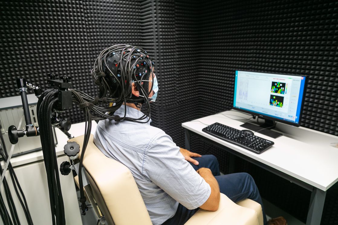
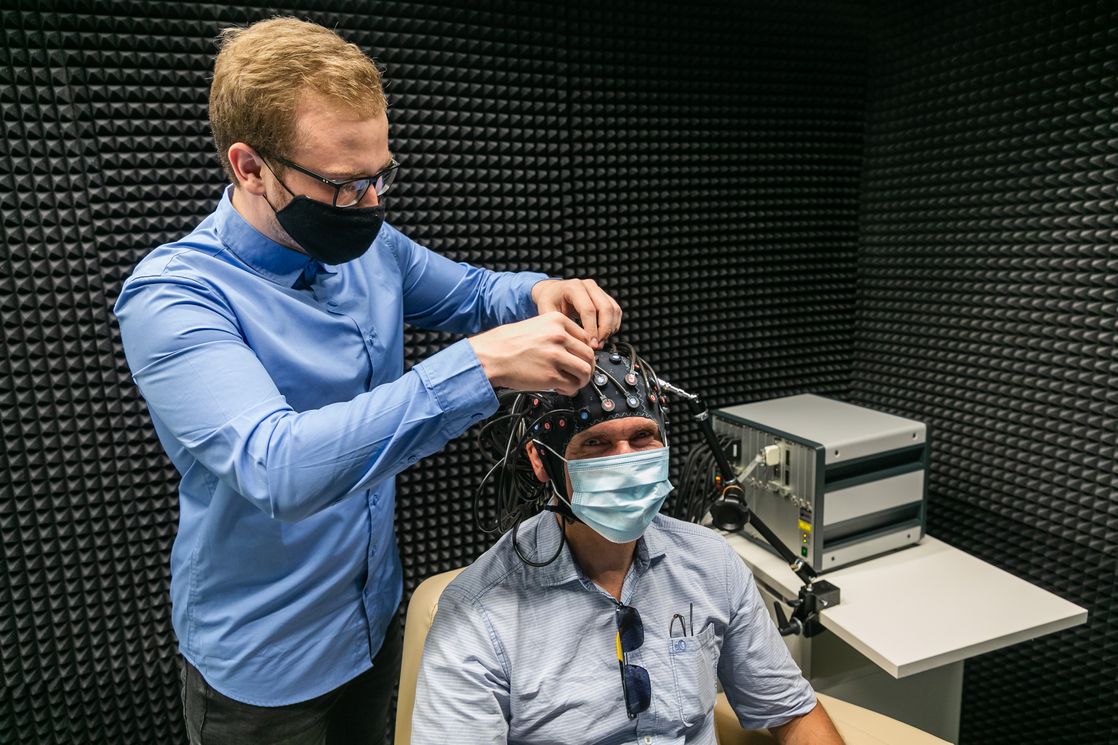
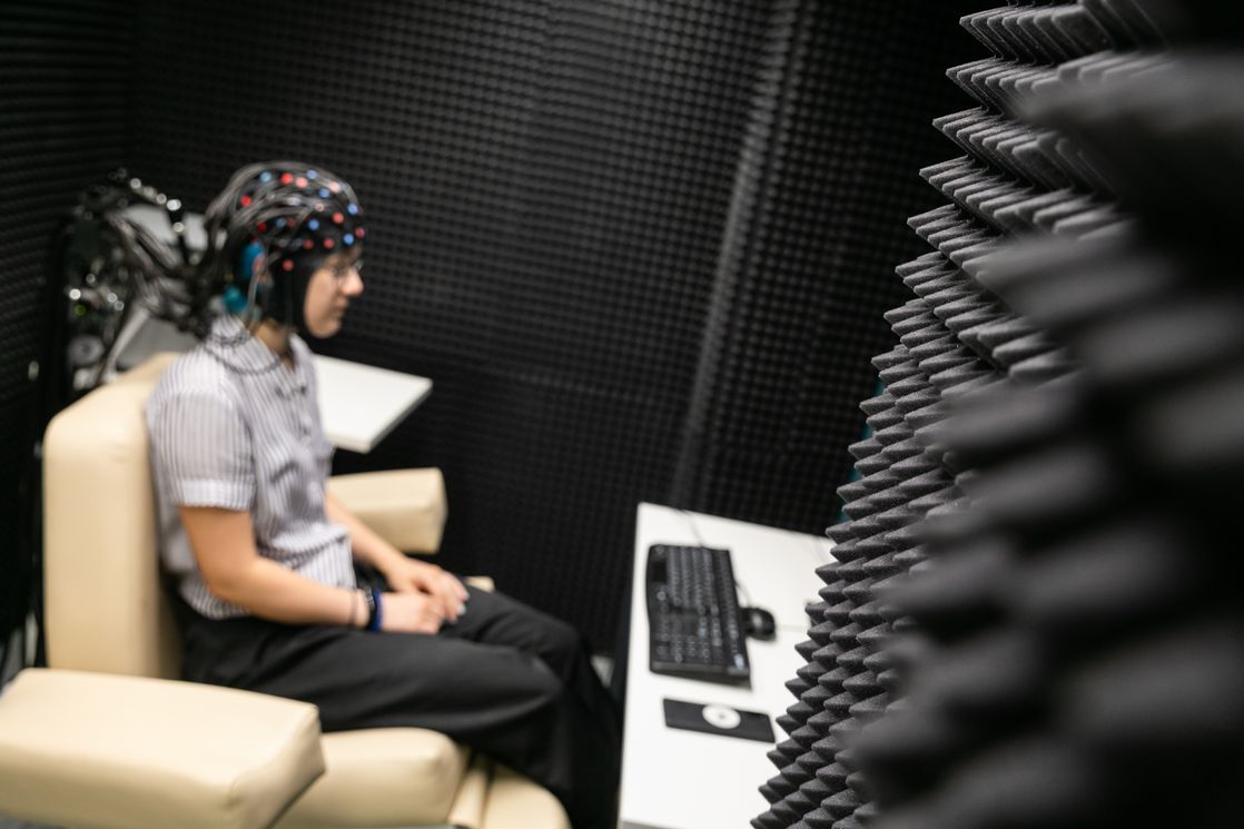
The Functional Optical Tomography laboratory looks like a medical room for EEG examinations. A comfortable chair stands next to a computer, which is connected to a ‘helmet’ made up of wires and dozens of sensors that envelop the test subject’s head. The room containing the optical tomograph itself is shielded by a ‘shell’ of special material that protects the device from external interference.
Aleksei Gorin, Junior Research Fellow at the Centre for Cognition and Decision Making at the ICN, told the HSE News Service how the process works. Special sensors conduct optical pulses from infrared lasers to detect differences in vascular tone in the veins and arteries. This in turn is used to measure oxygen absorption levels in various parts of the brain associated with logical problem solving and mental, sensory and motor activity. This allows researchers to study the brain's capabilities and its ability to respond to different tasks with more precision.
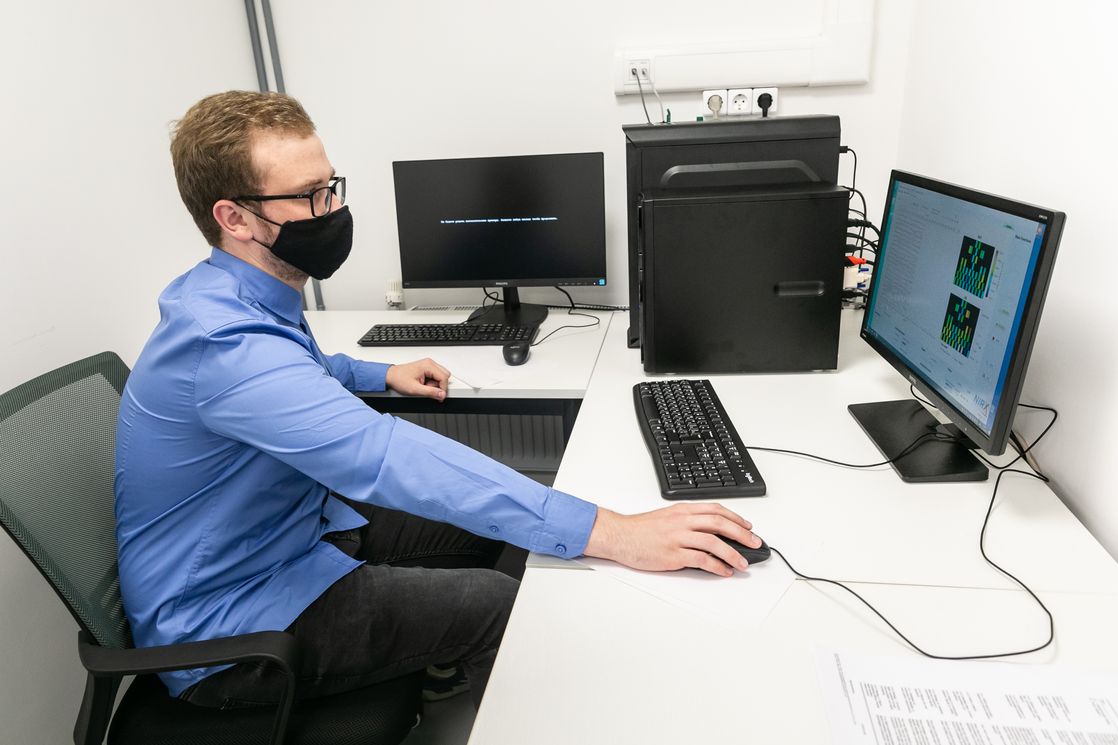
When our News Service reporter tested the equipment by reading a poem to himself, there was a drop in signal strength from the sensors monitoring the concentration of oxygenated blood. This, explained laboratory staff, reflects an increase in oxygen consumption in the brain.
The new equipment can also be used to study how the brain works when making certain decisions
The technology makes it possible to investigate certain kinds of brain activity and their impact on the brain's computational (and other) abilities. Optical tomography research also has medical applications, namely for the early diagnosis of heart and brain diseases.
In the absence of a perfect tool for studying the brain, a variety of experimental methods are required to get a clearer picture, Mr. Gorin explained. Researchers can conduct detailed behavioural research on the influence of various brain processes on human behaviour, and predict with a reasonable degree of accuracy which decisions an individual might make while affected by one process or another. ‘You can also give the brain a task and see how external factors affect how the brain works when performing decision-making tasks, sensory tasks or attention tasks. You can also learn more about how the brain works when someone is resting and relaxing’, he said.
Mr. Gorin also said that the Laboratory is interested in conducting interdisciplinary research with economists and medical professionals. Over the last two years, the Laboratory’s staff has expanded to include other specialists, including neurologists and psychiatrists. This has helped broaden the scope of its brain research.
Tatiana Chernyakova, Research Assistant at the International Laboratory of Social Neurobiology, said that the Laboratory’s plans for the near future include neuromarketing research and two cognitive experiments with the new equipment, using a combination of MRI and optical tomography to obtain new data.
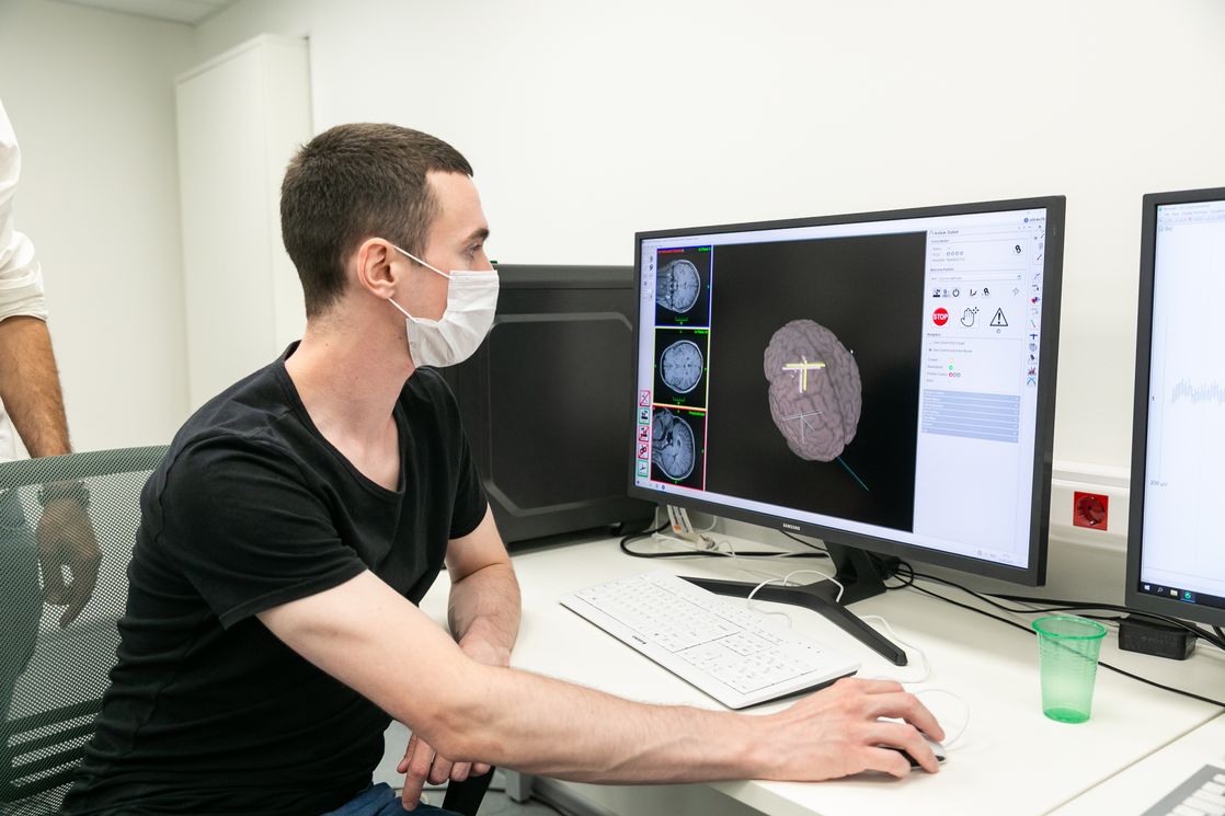
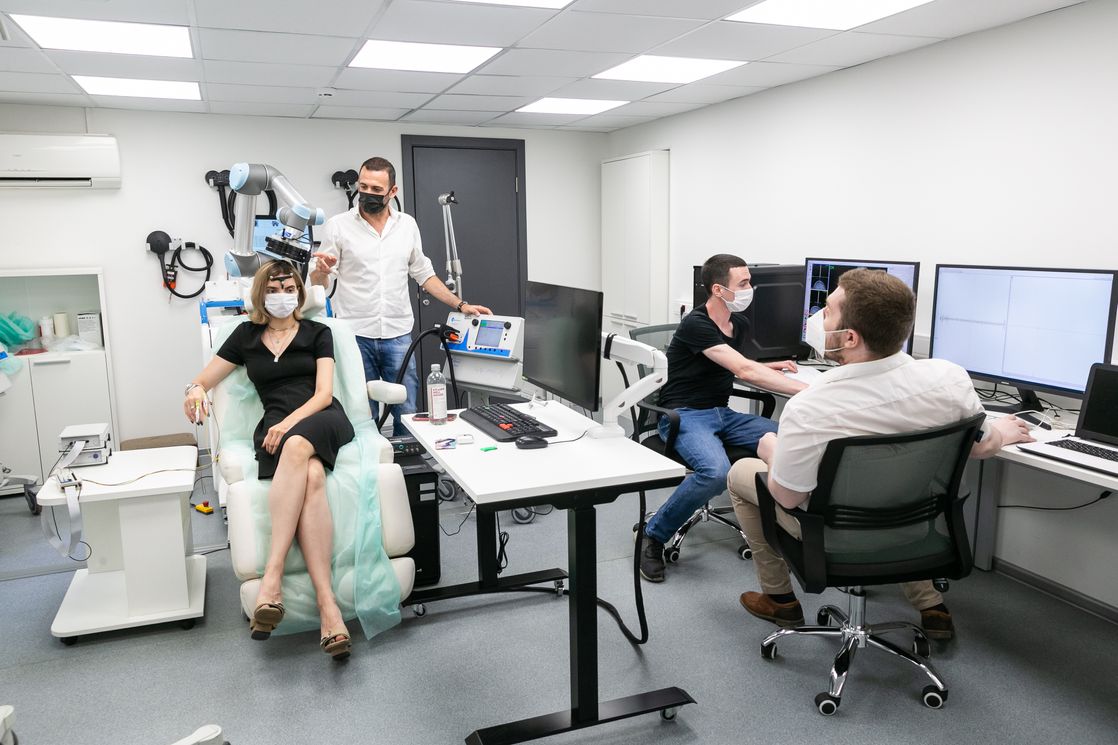
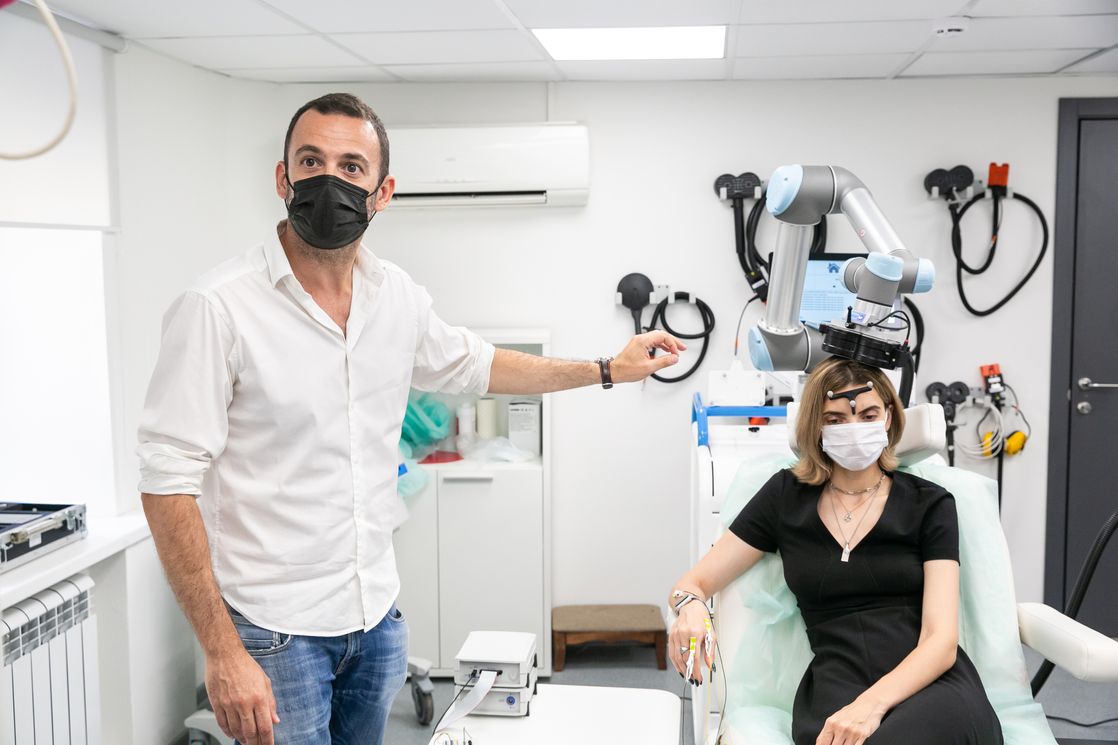
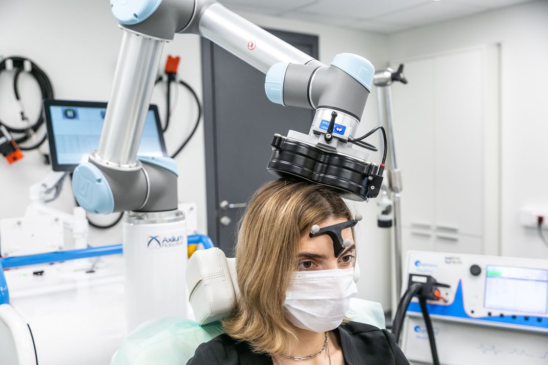
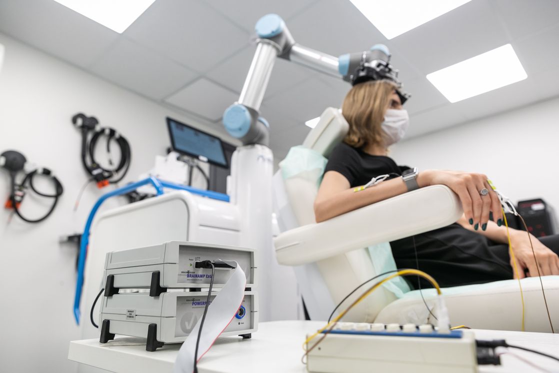
The Transcranial Magnetic Stimulation (TMS) laboratory has been overseeing the operation of a state-of-the-art stimulation device capable of activating parts of the brain. The device is installed next to a chair similar to one found in a dentist's office. A unique robotic arm (manufactured in France as there is currently no Russian equivalent) is placed above the head of a test subject and adapts to their movements. The accuracy of the robotic arm can be checked using a computer containing information about the area to be stimulated.
Matteo Feurra, Leading Research Fellow at the ICN Centre for Cognition and Decision Making, explains that if the subject makes any sudden movements for any reason (such as feeling unwell), the robotic arm deactivates the pulses and withdraws to a safe distance.
During the stimulation process, the robotic arm moves closer to the subject’s head, on which a reference bar has been placed to keep track of its position. The robot arm moves to match even the slightest movements of the head and sends an impulse to the target area of the brain. This induces the desired reaction, such as the contraction of muscles in the arm or fingers. While this result is visible in the test subject, the induced potential is also recorded and displayed by the computer.
Oksana Zinchenko, Research Fellow at the International Laboratory for Social Neurobiology, noted that stimulation has both research and medical applications. Once such use is the restoration of cells located around lesions or in the opposite hemisphere to an area affected by a stroke. Research Fellow Ainur Ragimova explained that transcranial magnetic stimulation can be used to treat depression and cervical dystonia, a disease that causes neck spasms, difficulty turning the neck and pain.
The TMS laboratory has a dedicated room for conducting complex research into brain stimulation and recording its effects. It is also shielded by a 'shell' for maximum signal clarity and isolation from external influences.
Areas of interest for this research include decision-making, social decisions, the speech areas of the brain, and memory. Ms. Zinchenko also explained that TMS is used to study the prefrontal cortex of the frontal lobe, which is responsible for selfish or prosocial motivations when deciding how to allocate resources—such as choosing whether or not to share points in a social game.
Using TMS to block the prefrontal cortex makes a person's behaviour more prosocial—they become more generous
Shutting down a group of neurons makes it possible to identify certain causal relationships. Researchers can see whether shutting down or stimulating individual areas disrupts or improves performance at a given task, and thus determine which areas of the brain are responsible for said tasks. These findings can then be used to build cognitive maps.
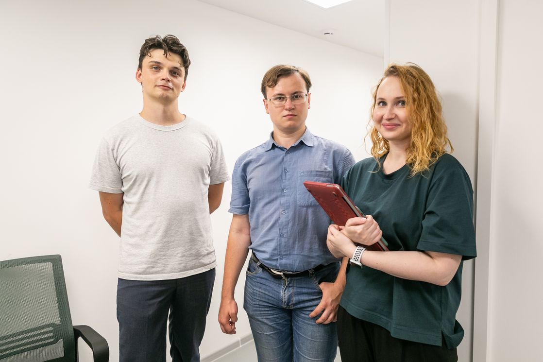
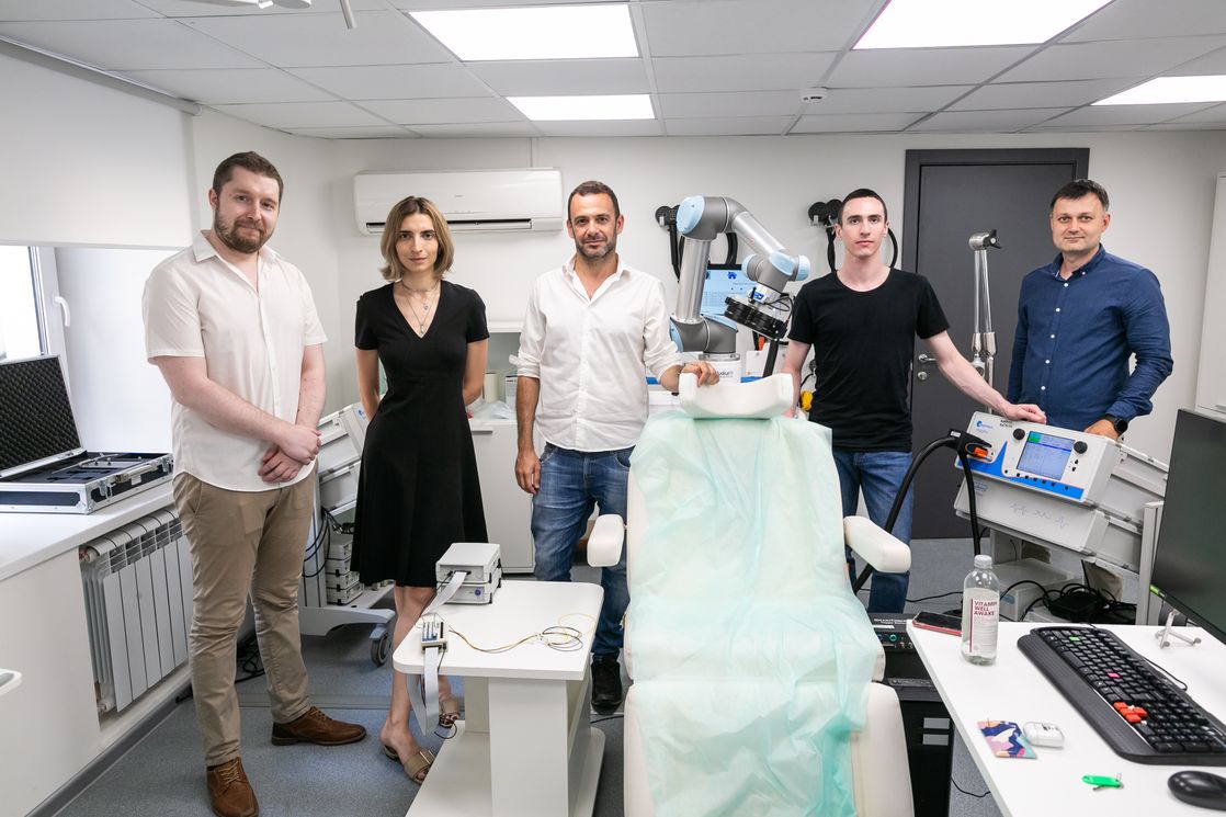
The laboratories and various research methods are available to specialists from different departments of the Institute for Cognitive Neurosciences, including foreign scientists such as Iiro Jääskeläinen, Academic Advisor of the International Laboratory of Social Neurobiology, Maria Del Carmen Herrojo-Ruiz, Leading Research Fellow at the ICN, Vladimir Djurdjevic, Research Fellow of the Centre for Cognition and Decision Making, Matteo Feurra and others.
Aleksei Gorin
Vladimir Djurdjevic
Tatiana Chernyakova
Maria Del Carmen Herrojo-Ruiz
See also:

Scientists Uncover Why Consumers Are Reluctant to Pay for Sugar-Free Products
Researchers at the HSE Institute for Cognitive Neuroscience have investigated how 'sugar-free' labelling affects consumers’ willingness to pay for such products. It was found that the label has little impact on the products’ appeal due to a trade-off between sweetness and healthiness: on the one hand, the label can deter consumers by implying an inferior taste, while on the other, it signals potential health benefits. The study findings have been published in Frontiers in Nutrition.
Internal Clock: How Heart Rate and Emotions Shape Our Perception of Time
Our perception of time depends on heart rate—this is the conclusion reached by neuroscientists at HSE University. In their experiment, volunteers watched short videos designed to evoke specific emotions and estimated each video's duration, while researchers recorded their heart activity using ECG. The study found that the slower a participant's heart rate, the shorter they perceived the video to be—especially when watching unpleasant content. The study has been published in Frontiers in Psychology.
Scientists Develop New Method to Detect Motor Disorders Using 3D Objects
Researchers at HSE University have developed a new methodological approach to studying motor planning and execution. By using 3D-printed objects and an infrared tracking system, they demonstrated that the brain initiates the planning process even before movement begins. This approach may eventually aid in the assessment and treatment of patients with neurodegenerative diseases such as Parkinson’s. The paper has been published in Frontiers in Human Neuroscience.
HSE Scientists Test New Method to Investigate Mechanisms of New Word Acquisition
Researchers at the HSE Centre for Language and Brain were among the first to use transcranial alternating current stimulation to investigate whether it can influence the acquisition of new words. Although the authors of the experiment have not yet found a link between brain stimulation and word acquisition, they believe that adjusting the stimulation parameters may yield different results in the future. The study has been published in Language, Cognition and Neuroscience.
'Our Goal Is Not to Determine Which Version Is Correct but to Explore the Variability'
The International Linguistic Convergence Laboratory at the HSE Faculty of Humanities studies the processes of convergence among languages spoken in regions with mixed, multiethnic populations. Research conducted by linguists at HSE University contributes to understanding the history of language development and explores how languages are perceived and used in multilingual environments. George Moroz, head of the laboratory, shares more details in an interview with the HSE News Service.
When Thoughts Become Movement: How Brain–Computer Interfaces Are Transforming Medicine and Daily Life
At the dawn of the 21st century, humans are increasingly becoming not just observers, but active participants in the technological revolution. Among the breakthroughs with the potential to change the lives of millions, brain–computer interfaces (BCIs)—systems that connect the brain to external devices—hold a special place. These technologies were the focal point of the spring International School ‘A New Generation of Neurointerfaces,’ which took place at HSE University.
‘It’s Thrilling to Have an Opportunity to Discuss Your Scientific Ideas with Interested People’
The International Laboratory of Dynamical Systems and Applications at HSE University–Nizhny Novgorod conducts in-depth theoretical and applied research, including the study of ocean waves, solar corona reconnections, volcanic phenomena, and ship stability. The lab’s researchers, who have received more than 20 significant research grants over the past five years, actively cooperate with Russian and international colleagues from China, Spain, the USA, the UK, Brazil, and other countries. Prof. Olga Pochinka spoke to the HSE News Service about the laboratory’s work.
How the Brain Responds to Prices: Scientists Discover Neural Marker for Price Perception
Russian scientists have discovered how the brain makes purchasing decisions. Using electroencephalography (EEG) and magnetoencephalography (MEG), researchers found that the brain responds almost instantly when a product's price deviates from expectations. This response engages brain regions involved in evaluating rewards and learning from past decisions. Thus, perceiving a product's value is not merely a conscious choice but also a function of automatic cognitive mechanisms. The results have been published in Frontiers in Human Neuroscience.
Electrical Brain Stimulation Helps Memorise New Words
A team of researchers at HSE University, in collaboration with scientists from Russian and foreign universities, has investigated the impact of electrical brain stimulation on learning new words. The experiment shows that direct current stimulation of language centres—Broca's and Wernicke's areas—can improve and speed up the memorisation of new words. The findings have been published in Neurobiology of Learning and Memory.
HSE Researchers Discover Simple and Reliable Way to Understand How People Perceive Taste
A team of scientists from the HSE Centre for Cognition & Decision Making has studied how food flavours affect brain activity, facial muscles, and emotions. Using near-infrared spectroscopy (fNIRS), they demonstrated that pleasant food activates brain areas associated with positive emotions, while neutral food stimulates regions linked to negative emotions and avoidance. This approach offers a simpler way to predict the market success of products and study eating disorders. The study was published in the journal Food Quality and Preference.